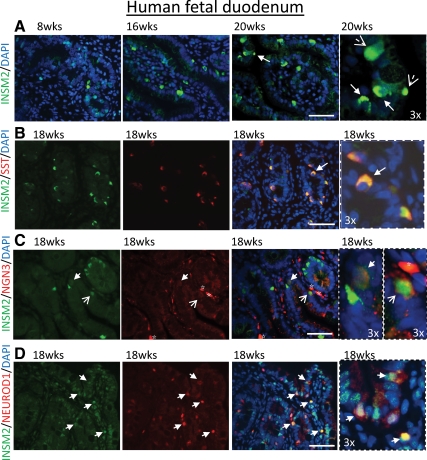
Fig. 5.
INSM2 in human fetal duodenum. A, Expression of INSM2 (green) in developing human duodenum (8–20 wk). B, INSM2 is colocalized with SST (red)-positive cells. INSM2 is coexpressed with NGN3 (C) or NEUROD1 (D) as indicated by arrows. Arrows (B–D) indicate INSM2+-costained cells. Images on fourth column are the amplification (×3) of the regions as indicated by the arrows on the third column. Nuclei were stained by 4′,6′-diamino-2-phenylindole (DAPI; blue). Scale bar: 50 μm.

