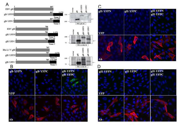
Figure 6. Generation and characterization of EBV and Rh-LCV BiFC constructs.
(A) Schematic diagram of EBV gB, EBV gH, and Rh gH with the N- and C-terminal fragments of Venus YFP and western blot analysis. CHO-K1 cells were transfected with EBV gB, EBV gH/gL, Rh gH/gL and the YFP-tagged constructs. Cell lysates were analyzed using the polyclonal gB antibody for EBV gB and gB-YFP constructs. CHO-K1 cells expressing EBV gH/gL, Rh gH/gL, and gH-YFP constructs were biotinylated as previously described and cell lysates analyzed using the HRP conjugated Avidin antibody. (B-D) Homologous glycoprotein interactions. (B) CHO-K1 cells were transfected with EBV gB-YFPN, EBV gB-YFPC, or both constructs together. Cells were fixed, permeabilized and stained with the polyclonal gB antibody and analyzed for gB expression (red channel) and YFP fluorescence (green channel) with DAPI staining of nuclei (blue channel). (C) CHO-K1 cells were transfected with EBV gH-YFPN, EBV gH-YFPC, or both constructs together with EBV gL. Cells were fixed, permeabilized and stained with the monoclonal E1D1 gH/gL antibody and analyzed for gH/gL expression (red channel) and YFP fluorescence (green channel). All confocal images were taken at 40X magnification. (D) CHO-K1 cells were transfected with Rh gHYFPN, Rh gH-YFPC, or both constructs together with Rh gL. Cells were fixed, permeabilized and stained with the monoclonal E1D1 gH/gL antibody and analyzed for gH/gL expression (red channel) and YFP fluorescence (green channel) with DAPI staining of nuclei (blue channel). All confocal images were taken at 40X magnification.

