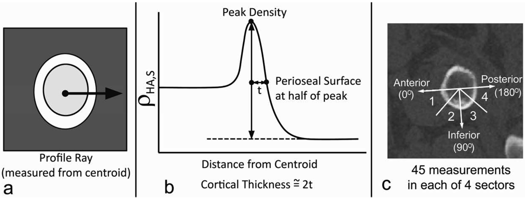
Figure 2.
Cortical thickness measurement method. a. The ρHA,S profile along a ray extending from the area centroid into the surrounding soft tissue was recorded. b. The distance between the location of the peak ρHA,S value and the location of the half-maximum on the periosteal margin was doubled to arrive at the cortical thickness measurement. c. A total of 45 measurements were made to compute the mean thickness in each of the sectors in the inferior half of the femoral neck section. Sectors were spaced in 45-degree increments starting with the principal axes in the anterior direction. The axes labeled in the figure correspond to anterior (0°) inferior (90°), and posterior (180°) directions.

