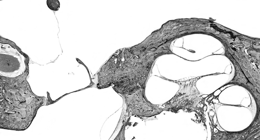
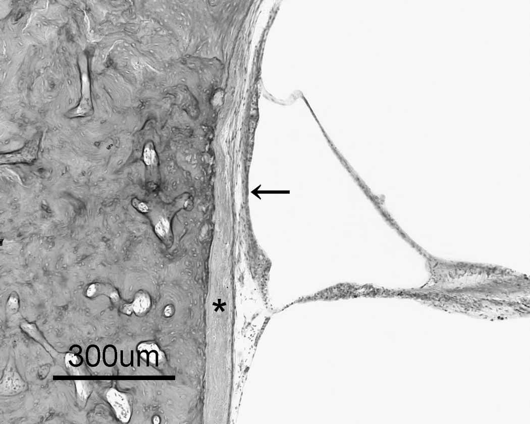
Figure 1.


a: Otosclerosis focus is involving cochlear endosteum and extending to the stapes footplate. 1b: A high magnification of the cochlear endosteum shows the hyalinization of the spiral ligament (*); and atrophy of the stria vascularis (arrow) (H&E staining).
