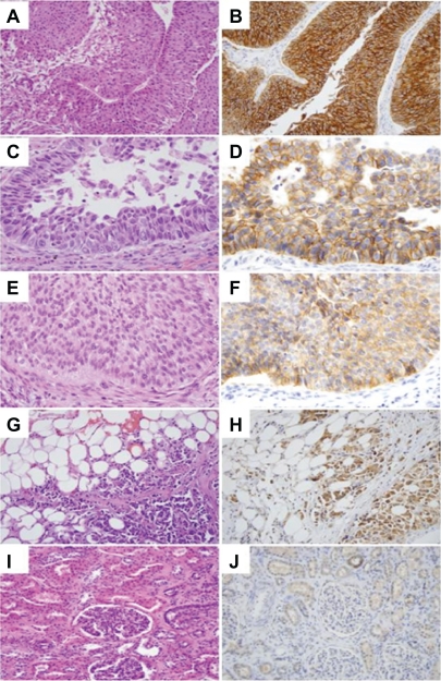
Figure 4.
FXYD3 immunohistostaining for UC of the renal pelvis. A, C, E, G, and I: H and E staining; B, D, F, H and J: FXYD3 staining. A, B) Low grade UC showing strong membranous and cytoplasmic staining. C, D) Area of high grade UC component showing strong membranous staining. E, F) Area of low grade UC component showing weak cytoplasmic staining. G, H) High grade UC cells invading perinephric fat showing strong FXYD3 staining. I, J) Normal appearing renal cortex showing no staining in kidney parenchyma including glomeruli and proximal tubules, while focal FXYD3 staining is occasionally seen in normal distal tubules.

