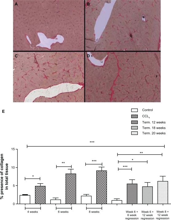
Figure 1.
Total collagen increase during fibrosis progression and regression.
The mean extent of total collagen found in liver sections, stained with Sirius red, from control, intralipid-treated animals (A); in CC l4-treated animals after 4 weeks of treatment (B); in CCl4-treated animals after 6 weeks of treatment (C); and CCl4-treated animals after 8 weeks of treatment (D). Quantification by Visiopharm software of the amount of total collagen expressed as a % of whole tissue in liver sections, showed a statistically significant increase in CCL4-treated rats compared with intralipid-treated animals at the same time of termination, for 4 weeks of treatment (P = 0.0100), 6 weeks (mean 8.25%, P = 0.0025), 8 weeks (P = 0.0007). Animals treated with CCl4 for 6 weeks and left to regress for another 6 showed a significant increase during regression in the total collagen content in the liver compared with the equivalent intralipid-treated animals at the same time of termination (P = 0.0079). Equally significantly increased levels of collagen were seen in both the groups undergoing 12 weeks regression after 6 weeks treatment (P = 0.0087), and 12 weeks’ regression after 8 weeks’ treatment (P = 0.0117). Quantitative histology revealed increased total collagen deposition during disease progression for animals treated with CCl4 for 4 weeks (mean 4.94% presence), 6 weeks (mean 8.25% presence) and 8 weeks (mean 9.11% presence). During regression, the total collagen deposition was gradually decreased to a mean of 6.9% (P = 0.0010) and 5.09% (P = 0.0117) for animals regressing for 6 and 12 weeks respectively after 6 weeks’ treatment and to a mean of 6.27% (P = 0.0083) in animals regressed for 12 weeks after 8 weeks treatment (E).

