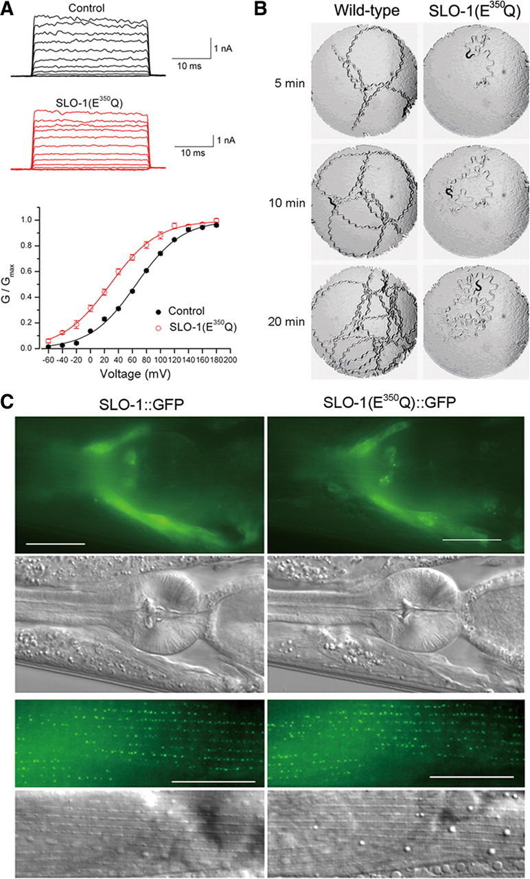
Figure 1.

SLO-1(E350Q) mutation shifted channel G–V relationship and inhibited worm locomotion. A, Macroscopic currents of inside-out patches from Xenopus oocytes expressing wild-type SLO-1 (Control) or SLO-1(E350Q) and the G–V relationship. The G–V relationship was fitted to the Boltzmann function G = Gmax/[1 + exp(V50 − V)/k], where G is the conductance at voltage V, Gmax is the maximal conductance, V50 is the voltage at which G = 0.5 Gmax, and k is the slope factor. The V50 was 65.4 ± 1.4 mV for wild-type SLO-1 (n = 10) and 28.4 ± 3.2 mV for SLO-1(E350Q) (n = 6). B, Locomotion behavior of a wild-type worm and a transgenic worm expressing Pslo-1::SLO-1(E350Q). A piece of paper with a circular hole (7 mm in diameter) was laid on top of the bacterial lawn in the agar plate to restrict animal movements within the field of observation. A single adult hermaphrodite was placed in the center of the field to start the assay. Snapshots of the animal and its locomotion track were taken at 5, 10, and 20 min. The animal expressing SLO-1(E350Q) showed a distorted locomotion waveform and greatly reduced locomotion velocity. C, SLO-1(E350Q)::GFP localized similarly as SLO-1::GFP in both neurons and body-wall muscle cells. The top two rows show fluorescent and differential interference contrast (DIC) images of the head. Fluorescent signal was localized mainly in the nerve ring region. The bottom two rows show fluorescent and DIC images of body-wall muscle. Fluorescent signal was enriched at dense body areas. Scale bars, 20 μm.
