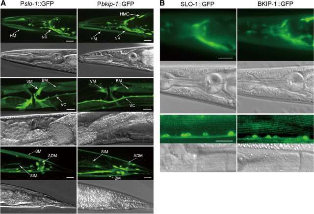
Figure 4.
Expression and subcellular localization patterns of bkip-1 were similar to those of slo-1. A, GFP showed similar expression patterns with Pslo-1 and Pbkip-1. Strong expression was observed in head neurons (not labeled), nerve ring (NR), ventral cord (VC), tail neurons (not labeled), body-wall muscle (BM), head muscle (HM), vulval muscle (VM), anal depressor muscle (ADM), and stomatointestinal muscle (SIM). Note that bkip-1, but not slo-1, was also expressed in head mesodermal cell (HMC). Because of the mosaic expression of the transgenes, fluorescence intensity was somewhat variable from cell to cell. B, Subcellular localization of BKIP-1::GFP and SLO-1::GFP fusion proteins in the nerve ring and body-wall muscle cells were similar. The green puncta in body-wall muscle cells correspond to locations of dense bodies. Scale bars, 20 μm.

