Figure 9.
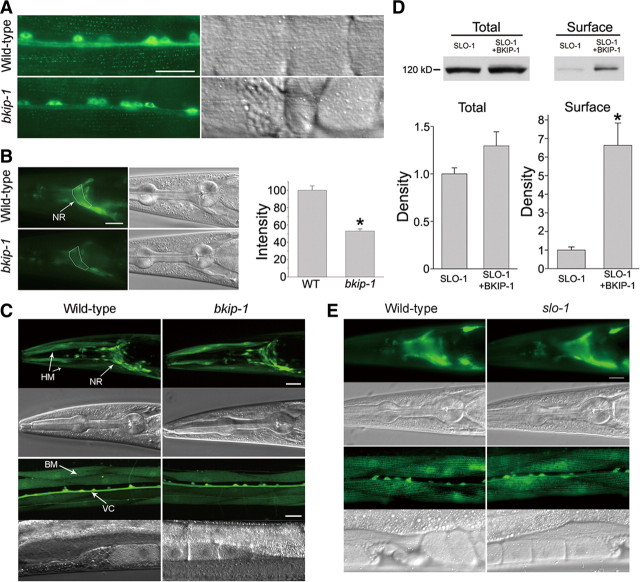
BKIP-1 increased SLO-1 expression in C. elegans and transfected HEK293 cells. A, SLO-1::GFP expression in body-wall muscle cells. SLO-1::GFP puncta at dense bodies appeared weaker in bkip-1(zw10) mutant compared with the wild type. The bright signal in the middle was of the ventral cord and motor neuron somas. B, SLO-1::GFP epifluorescence in the nerve ring was decreased in bkip-1(zw10) mutant. The mean fluorescence intensity of the nerve ring region (surrounded by the white line) was measured with NIH Image J software and compared between the two groups (n = 15 for both groups). * indicates a statistically significant difference compared with the wild type (WT) (p < 0.01, paired t test). C, slo-1 transcription was not altered by bkip-1(lf). GFP was expressed under the control of slo-1 promoter. GFP expression in neurons and muscle cells was indistinguishable between the wild type and bkip-1(zw10). NR, Nerve ring; VC, ventral cord; BM, body-wall muscle; HM, head muscle. D, Effects of BKIP-1 on SLO-1 total and surface protein levels. HEK293 cells were cotransfected with either SLO-1 and BKIP-1 or SLO-1 and the empty vector for BKIP-1. The amount of SLO-1 protein was quantified by densitometry and normalized first by α-tubulin and then by SLO-1 control (without BKIP-1). * indicates a statistically significant difference (p < 0.05, paired t test). Data are shown as mean ± SE and were from three experiments. E, BKIP-1 expression was not altered in slo-1 mutant. BKIP-1::GFP fusion was expressed in the wild-type and slo-1(md1745) mutant under the control of bkip-1 promoter. GFP epifluorescence in both neurons and body-wall muscle cells was indistinguishable between the wild-type and the slo-1 mutant. Scale bars, 20 μm.

