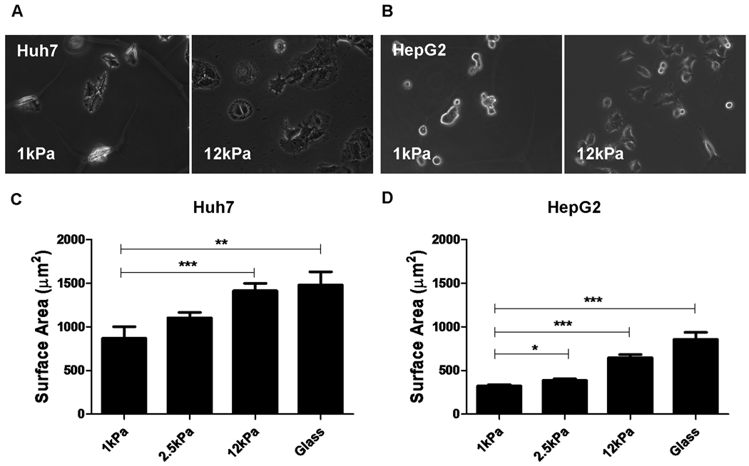
Figure 1. Changes in matrix stiffness regulate HCC cell morphology and spreading.
Huh7 and HepG2 cells were cultured on collagen-I-coated polyacrylamide gels with “tunable stiffness” (expressed as shear modulus, G’) in the range of 1–12kPa and collagen-I-coated glass. The stiffness values of the polyacrylamide gel supports used were selected in order to reflect range of stiffness values encountered in normal and fibrotic livers. Phase-contrast photomicrographs demonstrate the regulation of cellular morphology by support stiffness in both (A) Huh7 and (B) HepG2 cells. The surface area (square microns) of (C) Huh7 and (D) HepG2 cells was calculated by digital image analysis of phase-contrast images of cells on polyacrylamide gel supports. In each case, values reflect the mean (±SEM) of measurements from 50 cells in 3 independent experiments (*p<0.05, **p<0.01 and ***p<0.001).

