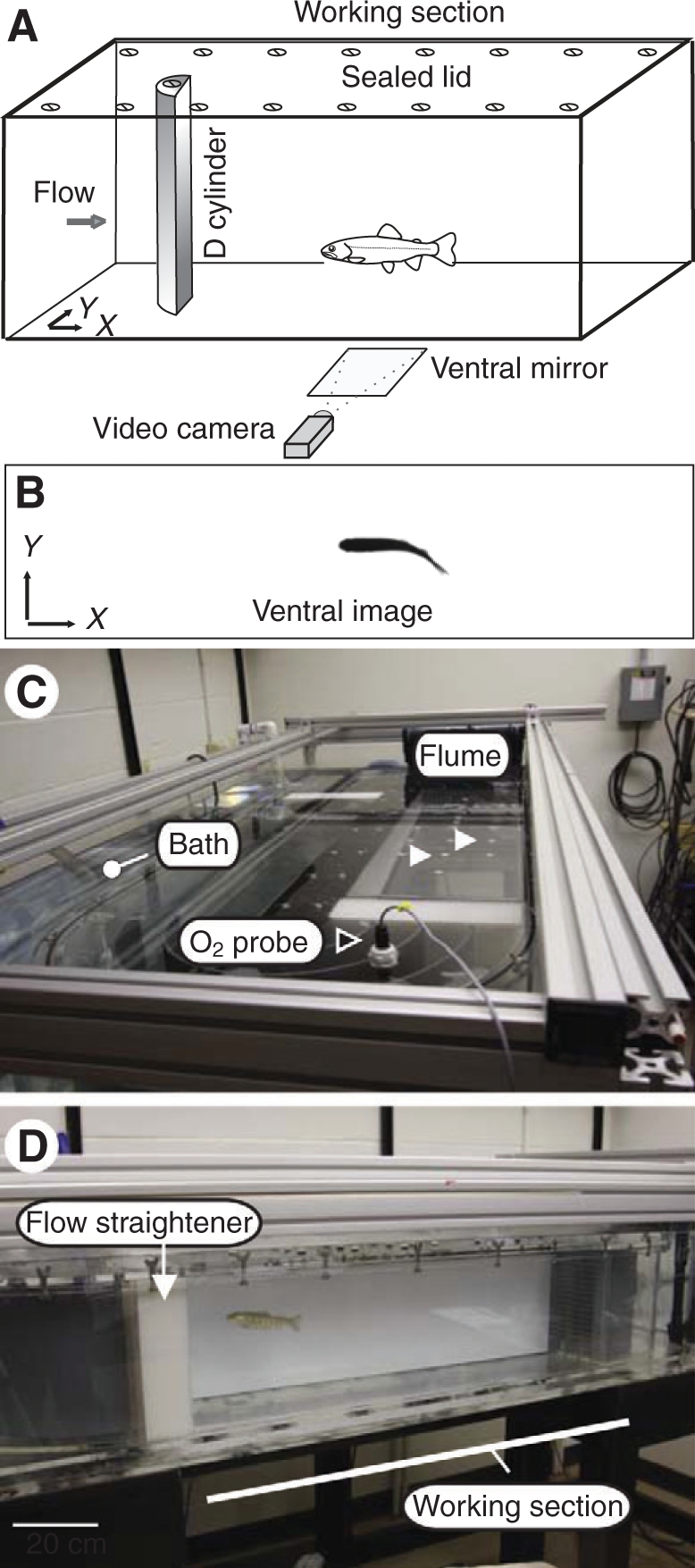
Fig. 1.

(A) Schematic of experimental setup. Fish were sealed in a flume respirometer in which a cylinder could be mounted. (B) A video camera pointed at a 45 deg front-surface mirror provided a ventral view of the position of the fish during each experiment. (C) Image of the flume respirometer illustrating the position of the oxygen probe (black arrowhead) and the drilled ports for cylinder placement (white arrowheads). The flume was submerged in an ambient temperature water bath (white dot) that is maintained at 100% oxygen saturation, which served as the source to flush the flume between experimental trials (see text). (D) Lateral view of the working section of the flume. Scale bar, 20 cm.
