Fig 6.
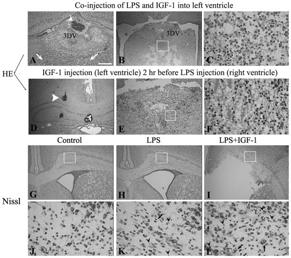
IGF-1 at a higher dose (5 μg/animal) enhanced LPS-induced brain injury. Co-injection of LPS with the higher dose of IGF-1 resulted in severe hemorrhage inside the third dorsal ventricle (3DV) (A, arrow head), and massive leukocytes infiltration (A, arrows) surrounding the 3DV. In some animals, the brain tissue around the 3DV (especially the ventral part) was severely damaged and totally replaced with infiltrated inflammatory cells (B, boxed area is highlighted in C). A separate injection of LPS with IGF-1 (injected 2 hr before LPS) resulted in similar pathology (brain hemorrhage shown in D, tissue loss shown in E and leukocyte infiltration shown in F). At P8, the Nissl-stained sections at the bregma level show enlarged lateral ventricle (H) and cell death (arrow heads in K) in LPS-treated rat brain, as compared to the control (G and J). IGF-1 deteriorates LPS-induced ventricle dilation (I). A marked increase in both pyknotic cells (arrow heads) and PMNs (arrows) was found in the LPS+IGF-1 treated rat brain white matter (L). Scale bar: in A, B, D, E, G, H&I: 200 μm; C, F, J, K&L: 50 μm.

