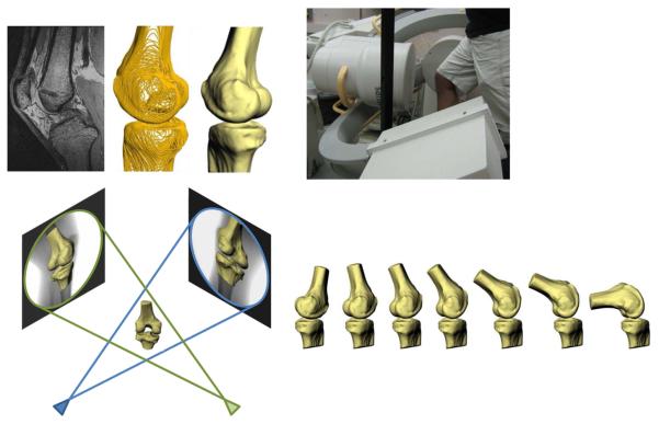
Figure 2.
High resolution MR images were segmented to create 3D models of the knee (top left). Next, the patients were imaged using biplanar fluoroscopy while performing a quasi-static lunge (top right). The fluoroscopic images and 3D models were then used to reproduce the motion of each subject's knee during the lunge (bottom).

