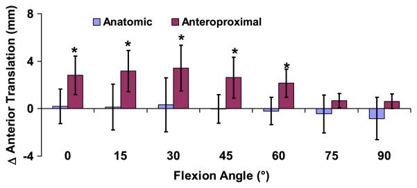
Figure 3.
The increase in anterior tibial translation of the reconstructed knee relative to the contralateral intact knee was measured as a function of flexion (mean and 95% confidence intervals). Zero denotes a knee that exactly mimics the motion of the contralateral side. Patients with grafts placed anteroproximally on the femur had increased anterior tibial translation relative to the contralateral side between 0 and 60° of flexion, while the anatomically placed grafts more closely restored normal knee motion. (*p < 0.05)

