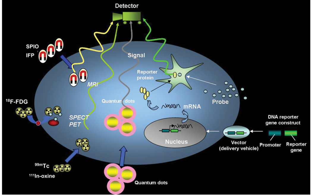
FIGURE 1.
Schematic for noninvasive imaging of stem cell fate in myocardium. Four different techniques include magnetic particle labeling, radionuclide labeling, quantum dot labeling, and reporter gene labeling. First 3 techniques are considered physical labeling, whereas last technique is considered genetic labeling. SPIO = superparamagnetic iron oxide; IFP = iron fluorescent particles. (Reprinted with permission of (45).)

