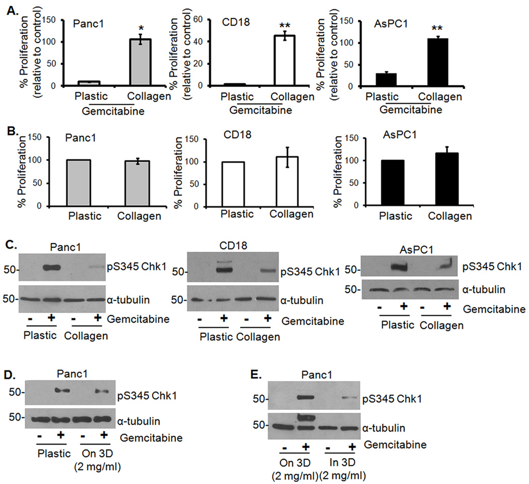
Figure 1. Growth in 3D type I collagen protects PDAC cells from gemcitabine-induced cell cycle arrest.
A. Panc1, CD18 and AsPC1 cells growing on plastic or in 3D type I collagen gels (2 mg/ml) were treated with gemcitabine (100 µM) for 24 hours. The effect on proliferation was quantified by determining the ability of cells to incorporate 3H-thymidine and normalized to control untreated samples. *, p < 0.05 relative to cells grown on plastic; **, p < 0.01 relative to cells grown on plastic. B. PDAC cells were plated on tissue culture plastic or in 3D I collagen gels for 24 hours. The effect on proliferation was quantified by 3H-thymidine incorporation. C. To examine the effect on checkpoint activation, cells were extracted out of collagen with collagenase treatment and the lysates immunoblotted for pS345Chk1 and α-tubulin. D, E. Panc1 cells were grown on plastic versus on 3D collagen gels (D), or on 3D collagen versus in 3D collagen gels (E), treated with gemcitabine for 24 hours and immunoblotted for pS345Chk1 and α-tubulin.

