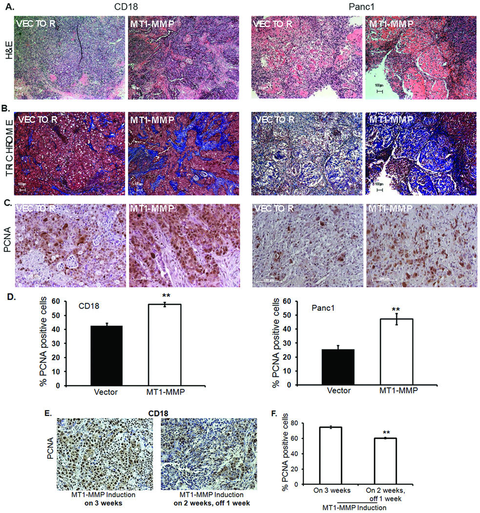
Figure 6. MT1-MMP attenuates gemcitabine-induced proliferation arrest in vivo.
Nude mice (n=5) were injected with Panc1 or CD18 cells expressing vector in the left flank and ΔC mutant of MT1-MMP in the right flank as detailed in Materials and Methods, and maintained on doxycycline water to induce MT1-MMP expression. Mice were injected with two doses of gemcitabine (15mg/kg) intraperitonially 2 days apart and the tumors excised and fixed in 10% formalin. Shown here are representative H&E (A), trichrome (B) and immunostaining for PCNA (C). D. Quantitation of PCNA (+) cells for CD18 and Panc1 was performed using Image J software. **, p <0.01 relative to cells expressing vector. E, F. Nude mice (n=10) were subcutaneously injected with CD18 cells expressing vector in the left flank and ΔC mutant of MT1-MMP in the right flank and maintained on doxycycline water for 2 weeks to induce MT1-MMP expression. Five mice were then switched to regular water for 1 week to block MT1-MMP induction. Four animals in each group were then injected with 2 doses of gemcitabine (15mg/kg) intraperitonially, 2 days apart; the tumors were excised and analyzed for the percentage of proliferating cells using PCNA staining and Image J software. **, p <0.01 relative to cells expressing MT1-MMP.

