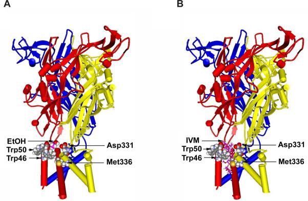
Fig. 2.
Molecular model of the rat P2X4R reveals a putative ethanol and IVM pocket. The model was built by threading the edited primary sequence onto the X-ray crystal structure of zebra fish P2X4R (Kawate et al., 2009). (A) A side view of the rat P2X4R showing the ectodomain and the six alpha helices of TM1 and TM2 segments of 3 different P2X4R subunits. Residues W46, W50 in the first alpha helix of one subunit as well as D331 and M336 in the final alpha helix of the adjacent subunit form a pocket that demonstrates a good fit for a molecule of ethanol (in pink) at the same scale. (B) A similar view of the rat P2X4R, but with a model of IVM (rendered in balls and sticks) inserted into a putative binding site in a position between the alpha helices like that described in nicotinic acetylcholine receptors (Sattelle et al., 2009). Figure taken from (Asatryan et al., 2010).

