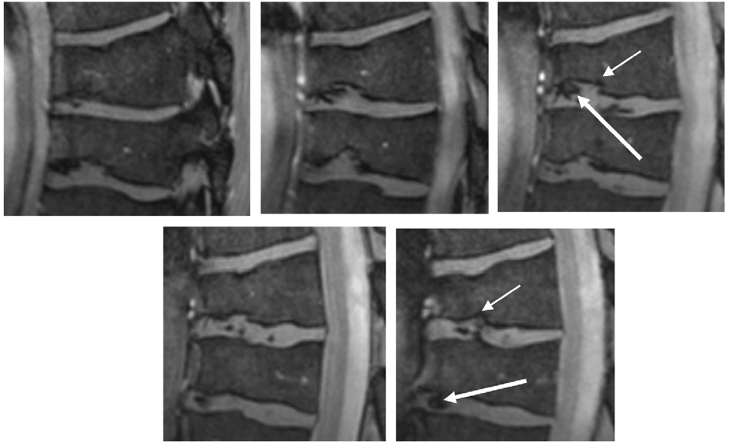
Fig. 4.
Zoomed images of two collapsed vertebrae from Fig. 2b. Cortical irregularity of the endplates can be seen (thin arrows), consistent with osteochondrosis. The inter-vertebral disks show inhomogeneities (thick arrows) which could be based on califications in the disk or, alternatively, degeneration.

