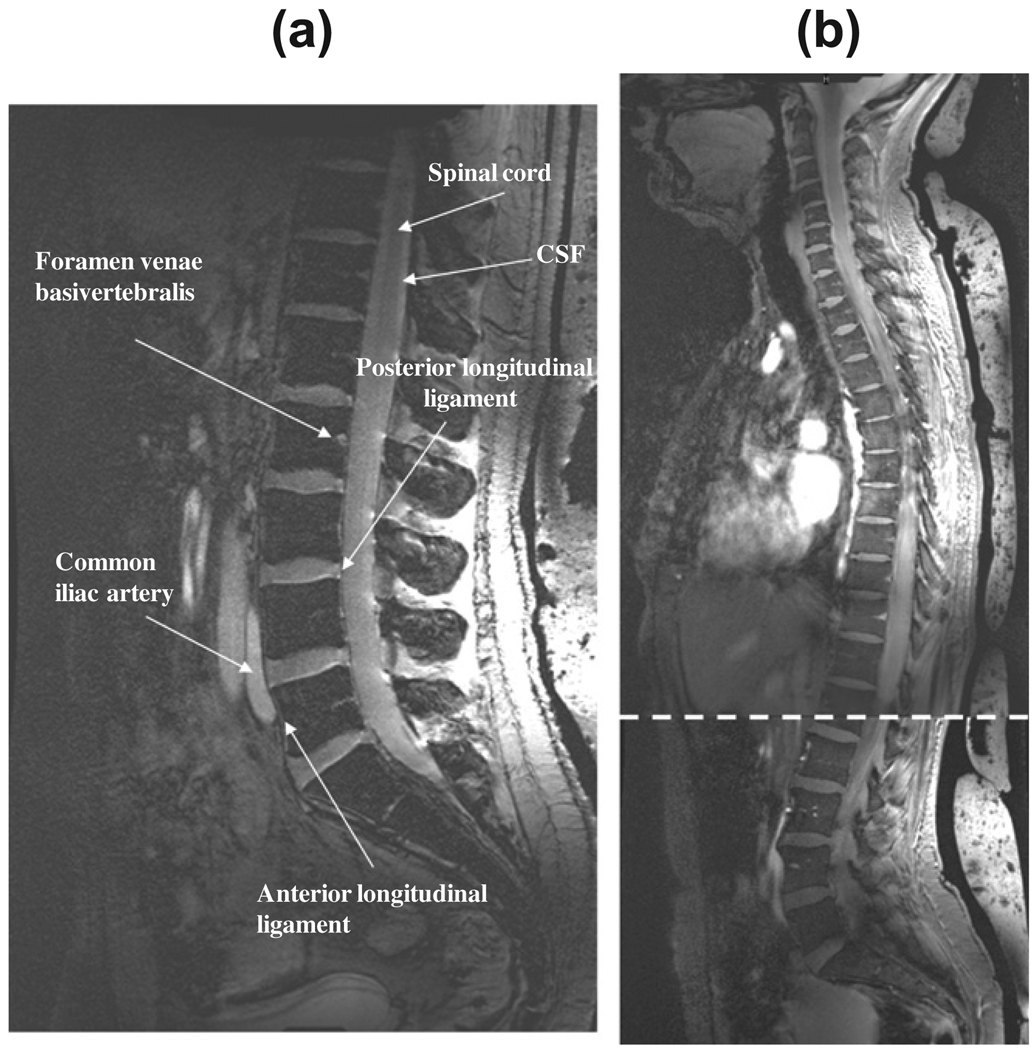
Fig. 5.
(a) A mid-sagittal image of the lumbar spine acquired from a patient, height 1.9 m, who weighted over 100 kg. (b) Two mid-sagittal slices of a female volunteer (height 1.7 m, 50 kg weight) which are acquired at two different positions of the patient table and stitched together at the overlap point indicated by the dotted line. No intensity correction or image smoothing has been applied to either image.

