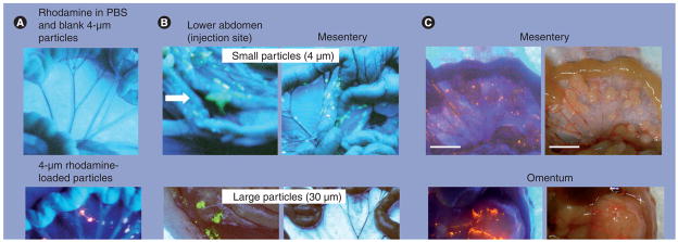
Figure 4. Intra-abdominal distribution of polymeric microparticles.
(A) Distribution. Tumor-free mice were given intraperitoneal (IP) injections of rhodamine dissolved in vehicle (0.01% Tween 80 in phosphate buffer solution) plus blank microparticles (top panel) or rhodamine-labeled microparticles (bottom panel). Rhodamine appears red under ultraviolet (UV) light. (B) Effect of particle size. Tumor-free mice were administered IP injections of acridine orange-labeled microparticles with average diameters of 4 or 30 μm. Acridine orange appears yellow under UV light. The smaller particles were dispersed throughout the cavity and on mesenteric membrane and omentum, which are common sites of local metastases of ovarian tumors. The larger particles were localized in the lower abdomen and were absent on mesenteric membrane and omentum. Arrows indicate the injection sites. (C) Localization of 4-μm particles on tumors. Mice were implanted with IP human ovarian SKOV3 xenograft tumors. After tumors were established (day 42), a mouse was administered an IP dose of rhodamine-labeled microparticles. At 3 days later, the animal was anesthetized and the abdominal cavity exposed. Photographs were taken in the region of the omentum and mesentery under UV light (left panels) and room light (right panels). Note the large tumor on the omentum (~13 mm [longest diameter]) and multiple small tumors on the mesenteric membrane (1–3 mm [longest diameter]). Red color under UV light indicated localization of rhodamine-labeled particles on the tumor surface. PBS: Plant-based solvent.
Reproduced with permission from [130].

