Figure 3.
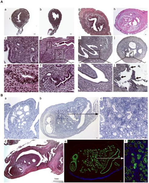
Histological analyses of uteri collected at different developmental stages from APCflox/flox and APCcko mice. Cross sections (Panel A, a & b) and longitudinal sections (Panel A, c & d) of 4wk old control (Panel A, a & c) and mutant (Panel A, b & d) uteri. Rectangular areas in Panel A, d are shown in higher magnification in e and f. Hyperplasia of epithelial lining of 5 month old APCcko mice uteri (Panel A, h) is shown compared with control (Panel A, g). (Panel A, i & k), polyp like outgrowths (Arrow) were observed from endometrium of some 7-month old mutant uteri (Panel A, j & l). Panel B, a shows development of carcinoma in situ in APCcko uteri. Panel B, b–d show mutant uteri with occlusion of uterine lumen by carcinogenic growth. Higher magnification image from Panel B, b showing admixed endometrial epithelial and stroma cells is shown in Panel B, c. Staining of mutant uterus with cytokeratin 8, an epithelial cell-specific marker. (Panel B, e). The boxed area is shown at higher magnification in Panel B, f. Bar equals 50 μm unless otherwise indicated.

