Fig. 1.
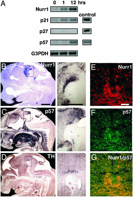
p57Kip2 expression is detected in differentiating DA cells in vitro and in vivo. (A) Dox-induced Nurr1 induces p21Cip1 and p57Kip2 mRNA expression. Nurr1, p21Cip1, p27Kip1, and p57Kip2 mRNA content was determined by RT-PCR in MN9D Nurr1Tet-On cells. cDNA-encoding p21Cip1, p27Kip1, and p57Kip2 were used as PCR controls. Sagittal sections of E13.5 wild-type mouse embryos showing Nurr1 (B), p57Kip2 (C), and TH (D) mRNA expression by in situ hybridization analysis. Images on the right are close-ups of the ventral midbrain. Nurr1 was expressed in a broad domain in the ventral midbrain as expected, whereas p57Kip2 mRNA was detected in a more restricted domain coinciding with TH mRNA expression. (E–G) Nurr1 (E) and p57Kip2 (F) immunoreactivity in confocal images of coronal midbrain sections. Overlay is shown in G. (Scale bars: B–D Insets, 100 μm; E–G, 50 μm.)

