
Fig. (5).
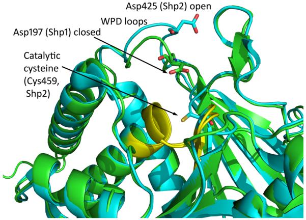
Overlay of the X-ray crystal structures of the PTP domain of Shp1 (pdb 1GWZ, colored green) and Shp2 (pdb 3B7O) colored blue, clearly indicating the closer proximity of the WPD aspartic acid residue to the catalytic cysteine residue for Shp1 as a consequence f the closed WPD loop conformation.