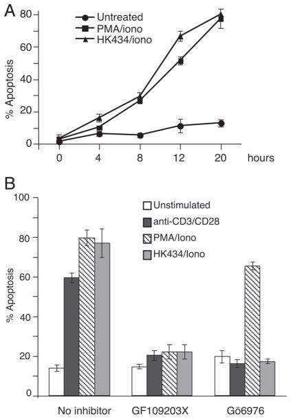
Figure 2.
Thymocyte apoptosis induced by DAG-lactone, HK434/ionomycin and PMA/ionomycin. (A) Thymocytes were stimulated with 1 μM DAG-lactone, HK434 or 0.26 ng/mL PMA with the time course indicated. Cells were stained for Annexin V and PI. Apoptotic cells are defined as Annexin V+ PI− cells. (B) Thymocytes were treated in the presence or absence of 1 μM Gö6976 or 1 μM GF109203X for 1 h followed by stimulation with 1 μM DAG-lactone, HK434 or 0.26 ng/mL PMA or 10 μg/mL anti-CD3/2 μg/mL CD28 (plate-bound). Twenty hours following stimulation, cells were stained for Annexin V and PI to assess apoptosis. Results shown in (A) and (B) represent four independent experiments with mean±SD indicated.

