Figure 1.

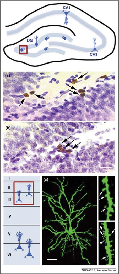
Parental experience produces changes in structural plasticity in the hippocampus and PFC. Top: Schematic diagram of the hippocampus showing the dentate gyrus, the location of adult neurogenesis (boxed area indicates region from which photomicrographs were obtained). Mother rats exhibit suppressed adult neurogenesis in the dentate gyrus prior to weaning of their offspring. (a) Virgin female rats have more proliferating cells, labeled here with the thymidine analog bromodeoxyuridine (BrdU) (arrows), compared with postpartum rats (b). BrdU-labeled cells are stained brown and cells labeled for Nissl are stained purple. Scale bar, 20 μm. Bottom: Schematic diagram of cortical layers (I–VI) in the PFC showing the neuron type affected by parenting (boxed area indicates layers II/III, where changes were detected in pyramidal neurons). Father marmosets exhibit enhanced dendritic spine density on pyramidal neurons of layer II/III PFC compared to non-fathers. (c) Layer II/III PFC pyramidal cell of a marmoset father labeled with the lipophilic tracer DiI (fluorescent green). Magnified views of DiI labeled dendritic segments showing dendritic spines (arrows) in a control (right upper) and a father (right lower). Scale bar, 30 μm for cell, 5 μm for dendritic segments. Adapted, with permission, from Ref. [25,73].
