
Figure 2. ES cells treated with XAV939 (XAV) from day 3 to 5 formed large areas consisting of spontaneously beating cardiomyocytes.
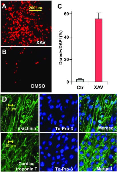
CGR-DsRed ES cells, which express DsRed-Nuc fluorescent protein under the control of cardiac α-MHC promoter, formed numerous red fluorescent nuclei following XAV treatment (A), versus relative few fluorescent nuclei following DMSO treatment (B). (C) Approximately 55.6% of DAPI+ nuclei co-expressed DsRed following XAV treatment, versus 1.8% following DMSO treatment (P=0.009). Results were obtained from 6 fields of XAV-treated EBs (on average, 59.6 DsRed+ cells out of 107.2 DAPI+ cells), and 6 fields of DMSO-treated controls (on average, 1.6 DsRed+ cells out of 92.2 DAPI+). (D) XAV939 treated ES cells formed larger areas of cardiomyocytes that expressed sarcomere proteins α-actinin (above) and cardiac Troponin-T (below). Left panels, immunostaining for sarcomere proteins (green). Middle panels, DAPI stained nuclei (blue). Right panels, merged images. Confocal images were taken using a Zeiss inverted LSM 510 confocal microscope (40×).