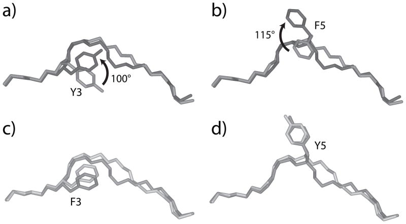
Figure 4.
Side chain conformational changes occur in A6 recognition of HuD but not Tax. A) Superimposed HuD peptides from the HuD/HLA-A2 and A6-HuD/HLA-A2 complexes, showing the 100° rotation of Tyr3 that occurs upon TCR binding. The peptide from the pMHC complex is light grey, the peptide from the TCR-pMHC complex is dark grey, with the arrow indicating the direction of rotation. B) Same as in panel A, but showing the 115° rotation in Phe5 that occurs upon TCR binding. C-D) Superimposed Tax peptides from the Tax/HLA-A2 and A6-Tax/HLA-A2 complexes, showing that unlike HuD, rotations in the positions of Phe3 (C) and Tyr5 (D) are not needed for TCR recognition. Peptides from the pMHC complex are light grey, peptides from the TCR-pMHC complex are dark grey.

