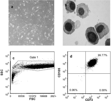
Fig. 1.
Photomicrographs of a plastic adherent human MSC (phase contrast, 200x) and b Giemsa-stained cytospin of human MSC (1,000x). c Forward and side scatter fluorescence-activated cell sorting (FACS) dot plot of human MSC. d Flow cytometry staining of passage human MSC showing positive staining for CD73 and CD105. CD45+ cells (leukocytes) constituted 0.4% at this time point.

