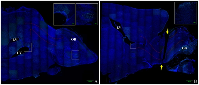
Figure 1. Mouse brain composite image illustrating olfactory bulb transection.
Images were obtained on a Nikon confocal microscope using a 20× objective and reconstructed into a composite image using ImageJ MosaicJ. DAPI labels cell nuclei and is labeled blue, anti-BrdU labels proliferating cells and is labeled green. Figure 1A is a representative image of a control (non-transected) brain, Figure 1B is a representative image of a transected brain. Approximate location of the rostral migratory stream (RMS) is outlined between the solid red lines, lateral ventricles are labeled LV, olfactory bulb is labeled OB, and the ends of the plane of transection are labeled with yellow arrows.

