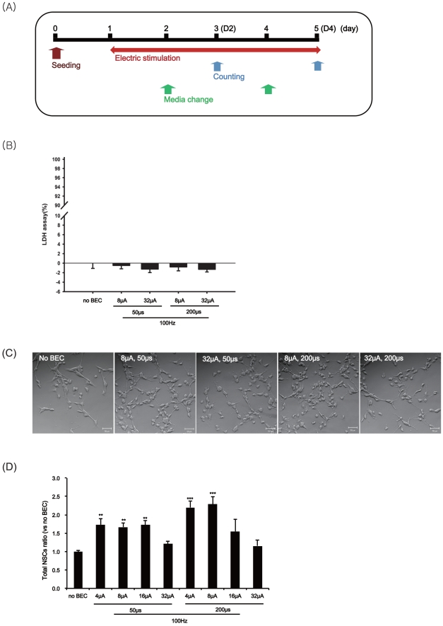
Figure 3. The effects of BEC on the proliferation of fetal NSCs.
The stem cells were stimulated at 100 Hz with a magnitude of 4, 8, 16 or 32 µA/cm2 and duration of 50 or 200 µs for 4 days and then counted the numbers of stem cells. (A) Experimental scheme with the electrical stimulation during the proliferation. (B) Cytotoxicity of BEC in fetal NSCs using LDH assay. Values are expressed as mean ± SEM of five to eight independent experiments. Data bars are not significantly different from No BEC group by ANOVA with Turkey test. (C) A representative phase contrast micrograph of fetal NSCs produced with each condition of BEC for 4 days; No BEC, 8 µA/cm2 for 50 µs, 8 µA/cm2 for 200 µs, 32 µA/cm2 for 50 µs, 32 µA/cm2 for 200 µs. (D) After 4 days of the electrical stimulation, cell numbers were counted with trypan blue staining. Data represent mean ± S.E.M. of n = 4–17. **, p≤0.01, ***, p≤0.001; by One-Way ANOVA: Tukey's HSD Post Hoc test.

