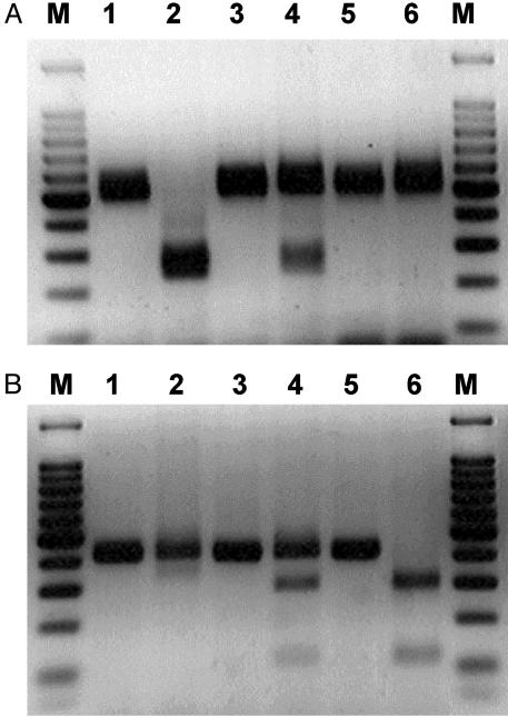
Fig. 2.
Detection of H67D and C294Y point mutations in the murine Hfe gene by genomic PCR amplification and subsequent BspHI and TaqI restriction site digestion. The H67D (CAT to GAT) destroys a BspHI site and C294Y (TGT to TAT) produces a TaqI new restriction site. Restriction enzyme analysis for each mutation introduced was performed by using homozygous, heterozygous, and normal control mice. (A) H67D mutation. Lane 1, undigested amplified PCR product from a normal control (500 bp); lane 2, DNA digested with BspHI from a normal control (240 and 260 bp); lane 3, undigested amplified PCR product from a heterozygote (500 bp); lane 4, DNA digested with TaqI from a heterozygote (240 bp, 260 bp, and 500 bp); lane 5, undigested amplified PCR product from a H67D homozygote (500 bp); lane 6, DNA digested with BspHI from a homozygote (500 bp). (B) C294Y mutation. Lane 1, undigested amplified PCR product from a normal control (417 bp); lane 2, DNA digested with TaqI from a normal control (417 bp); lane 3, undigested amplified PCR product from a heterozygote (417 bp); lane 4, DNA digested with TaqI from a heterozygote (128 bp, 289 bp, and 417 bp); lane 5, undigested amplified PCR product from a C294Y homozygote (417 bp); lane 6, DNA digested with TaqI from a homozygote (128 bp and 289 bp). The 100-bp markers were run at both ends of each gel.

