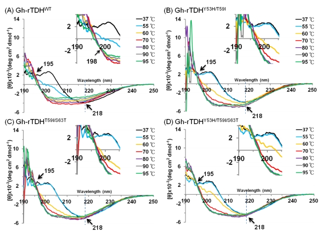
Fig 6.
Temperature dependence of the far-UV CD spectra of (A) Gh-rTDHWT, (B) Gh-rTDHY53H/T59I, (C) Gh-rTDHY53H/T59I and (D) Gh-rTDHY53H/T59I/S63T proteins in 10 mM phosphate buffer (pH 7.0) at 37, 55, 60, 70 oC. The arrows at 195 and 218 nm indicate the isodichroic points and the negative maximal peak, respectively. The arrow at 198 nm within the inset shows the second isodichroic point of Gh-rTDHWT protein.

