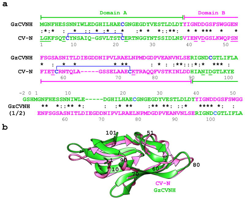
Figure 1. Sequence alignment and superposition of solution structures of CV-N from Nostoc ellipsosporum and GzCVNH from Gibberella zeae.
(a) Amino acid sequences of CV-N and GzCVNH. Domains A and B and the corresponding sequences are colored green and pink, respectively. The top sequence alignment is for CV-N and GzCVNH and the bottom compares the first and second sequence repeats of GzCVNH. Disulfide bonds are indicated by brackets and residues involved in protein-carbohydrate interactions are underlined in the CV-N sequence. Identical amino acids in the aligned sequence repeats are starred and conservative substitutions are marked by dashes. (b) Ribbon superposition of the NMR solution structures of GzCVNH (green) and wild type CV-N (pink). Amino acid positions for CV-N are indicated by numbers.

