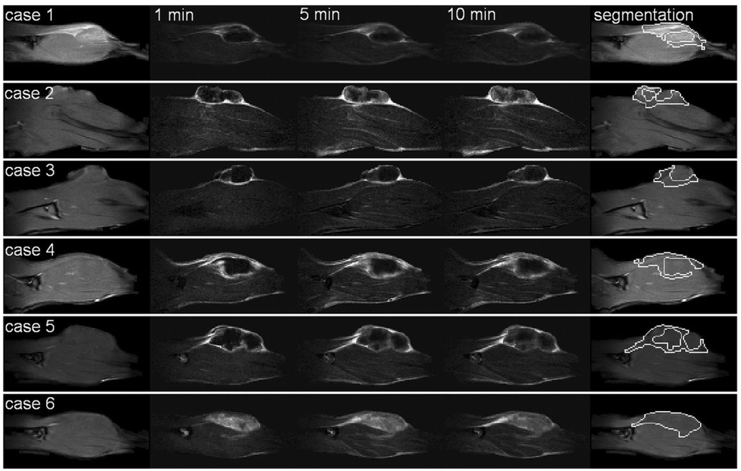
FIG. 1.
For six cases (rows), from left to right: the precontrast image, subtraction image at 1 min, subtraction image at 5 min, subtraction image at 10 min, and the viable tumor segmentation. The in-plane resolution is 0.31 mm × 0.31 mm. Images were cropped to a height of ~2.1 cm for display purpose. Except for case 6, all tumors developed a necrotic core.

