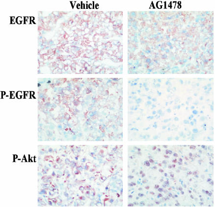
Fig. 2.
Immunohistochemistry analysis of U87MG.Δ2–7 xenografts treated with AG1478. U87MG.Δ2–7 xenografts were collected 30 min after injection of vehicle (Left) or AG1478 (1,000 μg) (Right) as described in Fig. 1. Frozen sections were cut and stained for expression of total EGFR, phosphorylated EGFR, and phosphorylated Akt.

