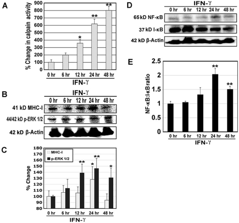
Fig. 2.

Changes in calpain activity and expression of MHC-I, p-ERK 1/2, NF-κB, and I-κB in rat L6 myoblast cells following IFN-γ stimulation. Cells were treated with IFN-γ (500 units/ml) for 6, 12, 24 and 48 hr. (A) Determination of percent changes in calpain activity (n = 6). (B) Representative Western blots to show levels of 41 kD MHC-I, 44/42 kD p-ERK 1/2, and 42 kD β-actin. (C) Determination of percent changes in 41 kD MHC-I (n = 5) and 44/42 kD p-ERK 1/2 (n = 3) expression based on Western blotting. (D) Representative Western blots to show levels of 65 kD NF-κB, 37 kD I-κB, and 42 kD β-actin. (E) Determination of NF-κB:I-κB ratio based on Western blotting (n = 3).
