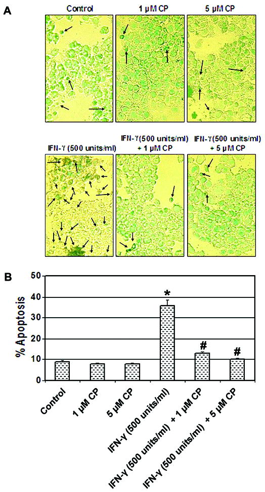
Fig. 3.

Attenuation of biochemical features of apoptosis by the calpain inhibitor. Cells were treated with IFN-γ (500 units/ml) for 24 hr. Calpeptin (1 and 5 μM) was added 5 min after the IFN-γ addition. (A) Photomicrographs showing representative cells from each treatment following ApopTag assay. The arrows indicate apoptotic cells. (B) Determination of percentage of apoptosis based on ApopTag assay (n = 3).
