
Figure 3. Examples of Activations in clinical models of pain.
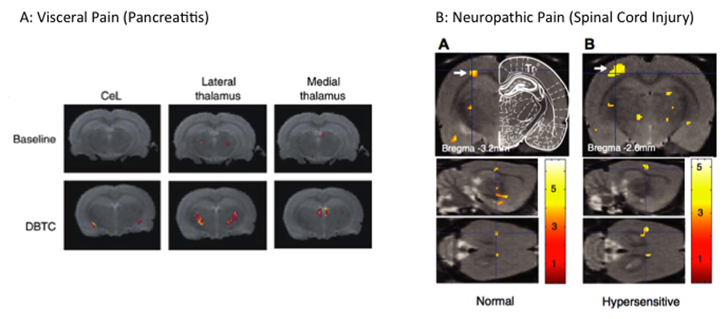
A: Visceral Pain: Activation in the thalamus in response to abdominal stimulation in a model of visceral pain ((Westlund et al., 2009); with permission, Neuroimage). Note the increase in the lateral and dorsal thalamus suggesting increased activation in lateral and medial pain pathways.
B: Neuropathic Pain: Activation in response to trunk stimulation in a rat model of spinal cord injury. Panel A shows activation in response to the stimuli in SI (arrow) in normal animals that is increased in the injured animal ((Endo et al., 2008a); with Permission, IASP Press).