Fig. 1.
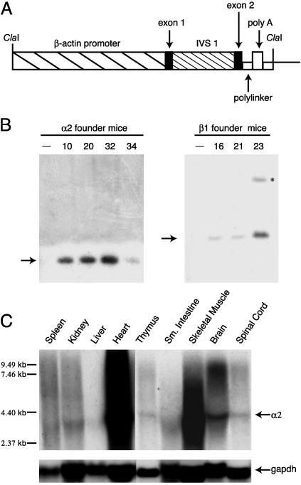
Generation and characterization of human α2 and β1 integrin transgenic mice. (A) Schematic diagram of plasmid pBAP consisting of 3 kb of human β-actin regulatory sequence, noncoding exon 1, intron 1, exon 2, and a polylinker into which α2 and β1 cDNAs were inserted. The polyadenylation signal is from simian virus 40. IVS, intervening sequence. (B) Southern blot hybridization analysis of genomic DNA from α2 (Left) and β1 (Right) transgenic founder mice. The hybridization probe was a HindIII fragment of α2 DNA (Left)ora BamHI fragment of β1 DNA (Right). Transgenic founder number is at top of each lane. Nontransgenic litter mates designated by -. Arrows indicate DNA fragments detected by α2 or β1 probes. Asterisk indicates incomplete digestion product. (C) Northern blot hybridization analysis of human α2 mRNA from mouse tissues of founder #20 offspring. The hybridization probe was a32P-labeled α2 (Top) or gapdh (Bottom) cDNA fragment. Arrows to the right of blots indicate α2 and gapdh mRNA. Molecular weight markers are to the left of the blot.

