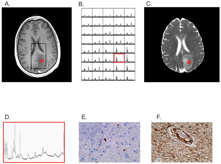
Figure 1.
Non-enhancing Grade 3 astrocytoma (A) contrast-enhanced T1-weighted MRI showing PRESS box (black) where MRS data was acquired and biopsy location (red). (B) 3D-MR spectra and (C) apparent diffusion coefficient image map were that were used to select the biopsy location prior to surgery. Post-surgical (F) HRMAS MR spectrum, immunohistochemical (D) Ki-67 and (E) VEGF labeling of biopsied tissue sample.

