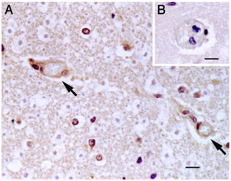
Fig. 4.
Immunohistochemical analysis of FKN expression in cerebral cortex of the patient with cerebral malaria. Excised tissues were stained with polyclonal anti-FKN-CD antibody (A) or goat IgG (B). Arrows indicate expression of FKN on EC of the microvasculature with sequestration. (Scale bar, 10 μM.)

