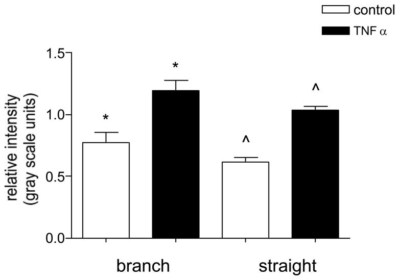
Figure 5. The expression of ICAM-1 is not different in straight and converging venular regions.
Control and TNFα activated venules were imunofluorescently labeled for ICAM-1 and the relative expression was quantified in straight venular regions (vessels 30–80μm diameter) and compared to regions of venular convergences (as defined in METHODS) located upstream in the same vessels. The expression of ICAM-1 in both regions was not significantly different in control or TNFα activated venules. */^ significantly different from each other (p<0.05), n=8 vessels.

