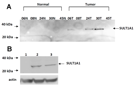
Figure 1.
Characterization of SULT1A1 expression in breast cancer tumors and cell lines. (A) Western blot analysis of SULT1A1 expression in matched specimens of infiltrating ductal carcinoma lysates (100 μg) and their corresponding normal breast tissue controls. (B) stably-transfected ER-dependent MCF7 cells, Lane 1, MCF7 pcDNA3 (50 μg), Lane 2, MCF7 SULT1A1 (50 μg), Lane 3, human liver cytosol (50 μg). Lysates were separated by SDS-polyacrylamide gel electrophoresis, blotted, and probed with anti-SULT1A1 antibody (1:1000) developed by Open Biosystems (Huntsviiie, AL) as described in the Materials & Methods.

