
Figure 1.
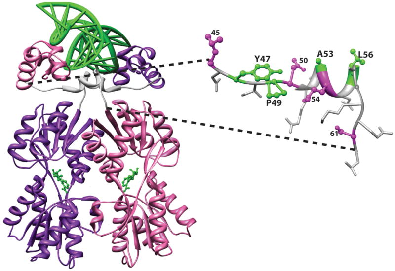
Representative structure of a LacI/GalR homolog with YPAL linker. The structure of the E. coli LacI dimeric variant from the pdb 1efa 39 was rendered with the program Chimera 58. On the left, the full length protein is shown with one monomer in purple and the other in pink. DNA is shown at the top of the figure as a green ladder; anti-inducer allosteric ligands are shown as green balls-and-sticks near the center of each monomer. On the structure of the full-length protein, the 18 amino acids that link the DNA-binding and regulatory domains are colored white. On the right side of the figure, the structure of one linker is expanded with linker side chains shown. YPAL positions 47, 49, 53, and 56 are colored green. A second group of positions with moderate conservation (45, 50, 54, and 61) are colored magenta. Nonconserved positions are shown as white sticks. Position 52 is the only side chain not visible; the side chain for position 57 (alanine in LacI) is near the base of the side chain for L56; the Cα of position 58 (glycine in LacI) is shown as a white ball on the backbone ribbon.