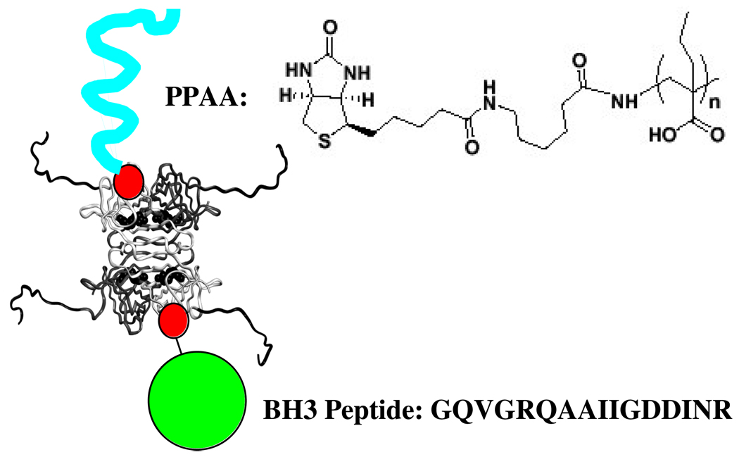
Figure 1.
Schematic representation of the TAT-SA complex with biotinylated PPAA and biotinylated BH3 peptide (biotin represented as red circle). The biotin linker and PPAA sequence are shown, together with the BH3 peptide sequence. The TAT peptide sequence has been modeled as a random coil extension onto the N-terminus of the four streptavidin subunits whose backbone folds are defined from x-ray crystallographic structures. They appear as the backbone extensions coming off the four subunits.

