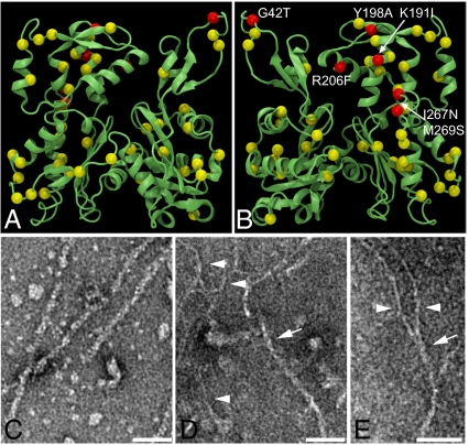
Fig. 1.
Divergent GiActin forms filaments. (A and B) Front and back views of giActin mapped on the F-actin structure with 48 nonconservative substitutions indicated in yellow and red. Nonconservative substitutions at filament contact points are in red. Note the substitutions in the DNaseI loop (residues 39–47) and the hydrophobic plug (residues 266–269). (C–E) TEM of negative-stained (C) rabbit actin and (D and E) giActin. Arrows point to ∼7-nm filaments; arrowhead points to ∼3.5-nm filaments. (Scale bar, 50 nm.)

