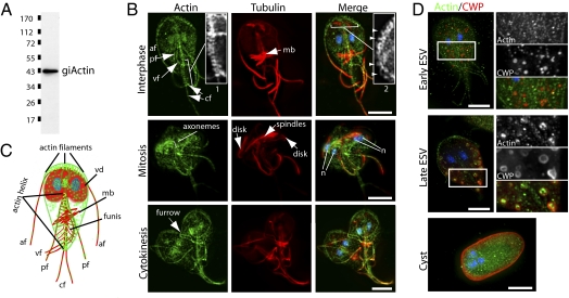
Fig. 2.
GiActin forms stage-specific structures. (A) Anti-giActin Western blot against total Giardia extract. (B) Immunofluorescence labeling of actin (green), tubulin (red), and DNA (blue) in trophozoites. Actin localizes to the cortex, the two nuclei, and all axonemes. An actin helix bundles the caudal flagella pair (cf) Inset 1, and short filaments are visible along the anterior flagella (af) Inset 2 (arrowheads). Note the repositioning of the af, ventral flagella (vf), and posterior flagella (pf) and enrichment of actin during mitosis. (C) Diagram of actin (green) and microtubule (red) cytoskeletons in trophozoites, including eight flagella, a ventral disk (vd) (parasite attachment), the median body (mb) (likely a microtubule reservoir), and the funis (rib-like microtubule structure). (D) Localization of actin (green) and cyst wall protein (CWP) (red) in encysting trophozoites. Note the recruitment of actin to mature encystation-specific vesicles ESVs. (Scale bar, 5 μm.)

