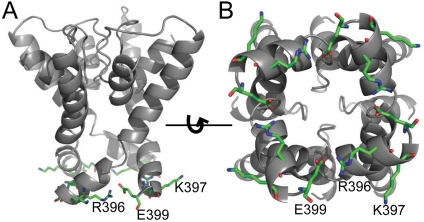
Fig. 6.
Structural model of the SK channel pore based on the Kv1.2 structure. Sequence of rSK2 was threaded into the structure of Kv1.2 using the “SwissModel” function in Swiss-PdbViewer software. (A). A view of the model from the side of the channel pore. R396, K397, and E399 are shown as sticks. Nitrogen atoms are shown in blue and oxygen atoms in red. (B). A view of the model from the intracellular side of the channel pore.

