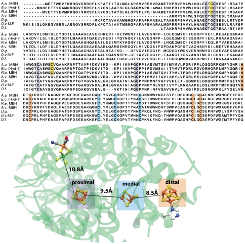
Fig. 1.
Top: Sequence alignment of O2-sensitive and O2-tolerant hydrogenases. A set of columns of the same color represents the conserved residues that bind a specific cluster, highlighted in the structure of D. vulgaris MF (1WUI pdb code). The two extra cysteines in the A. aeolicus enzyme are depicted in yellow. Bottom: Localization of the [NiFe] site and the three [FeS] clusters in the standard hydrogenases and the closest metal-to-metal distances between different cofactors. The [FeS] clusters are denoted as proximal (P), medial (M), and distal (D), based on their distance from the [NiFe] site (Fig.S1).

